Automated Knee Implant Recognition with Deep AI

Recognizes: Endotec — Buechel-Pappas

Identify knee replacement implant manufacturers and model systems from AP and Lateral X-ray images using AI-assisted image analysis with Grad-CAM visual explainability.
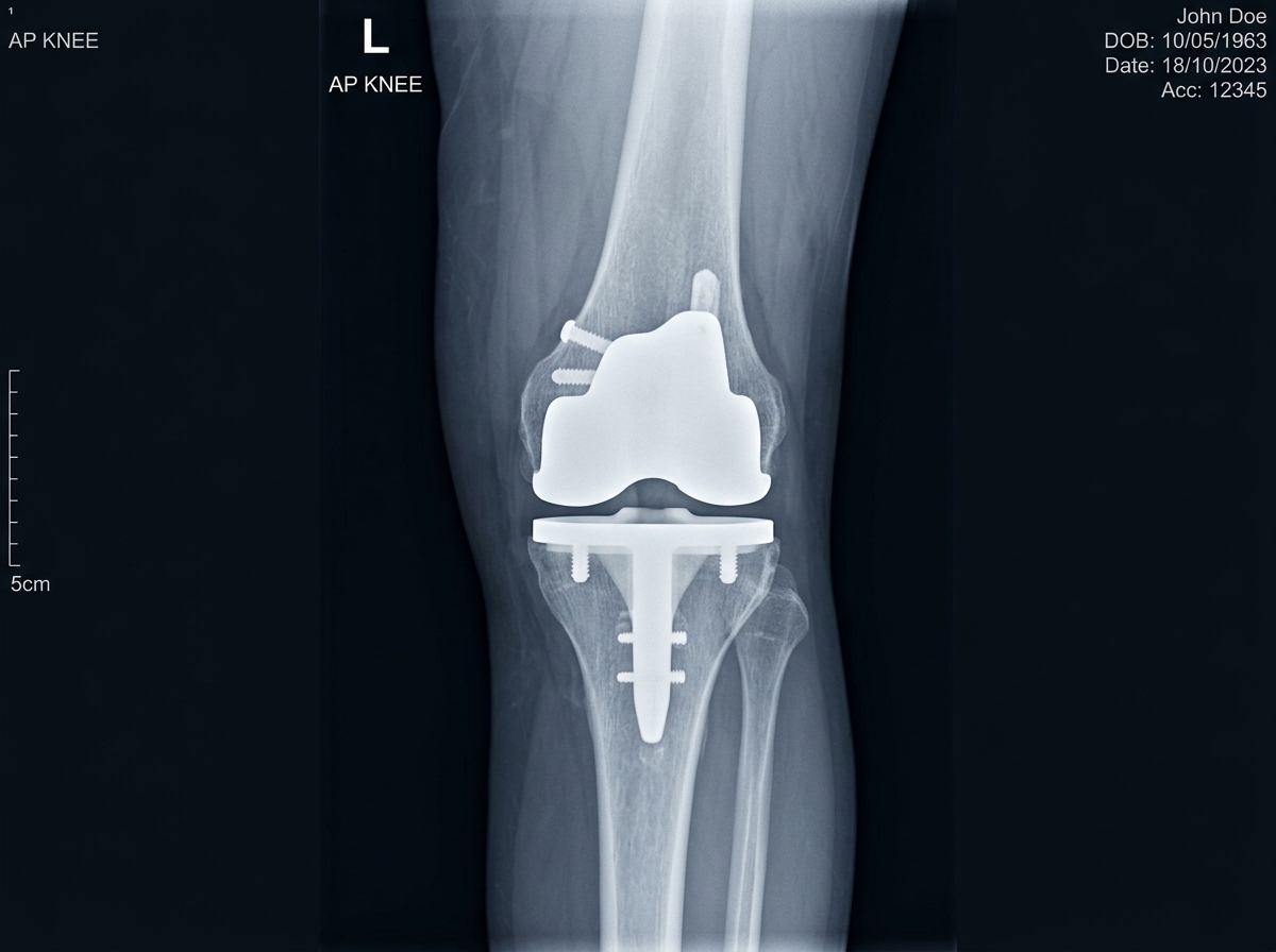
Live Scanner Demo
Sample #KNEE-8921
AI-generated illustration of a knee X-ray with a knee implant, not a patient image
AI-generated illustration — not a patient image
PRIMARY IDENTIFICATION
Stryker Triathlon Knee System
98.4% Confidence
Illustrative Example — Not a Validated Performance Metric

10 Systems

Supported Implant Classes

Native DICOM

16-bit VOI LUT Windowing

Top-3 Rankings

Multi-Class Softmax Confidence

Grad-CAM & ROI

Visual Heatmap Explainability

Automated Image Analysis

Orthopedic Implant Identification, Designed for Research

ImplantDetect processes standard knee radiographs to identify prosthesis manufacturers and specific component lines using AI-assisted visual feature analysis. Designed as an educational and pre-operative revision research tool, the system provides transparent candidate rankings paired with explainability overlays.

Top-3 Softmax Candidate Ranking Provides probability confidence scores across 10 major knee prosthesis systems.
Visual Feature Heatmap Overlay Grad-CAM gradient activation highlights condylar, stem, and tray feature regions.
Exportable PDF Reports & History Archive Archived securely to clinician accounts with structured PDF clinical summary exports.
Analysis Workflow Overview
01
Radiograph Upload & Preprocessing AP/Lateral views in PNG, JPG, WEBP, or raw 16-bit DICOM.
02
Pattern Recognition & Visual Heatmaps Deep learning inference produces top candidate probability & Grad-CAM ROI.
03
Clinical Documentation Export Download structured PDF case summary with timestamped audit metrics.

Supported Radiograph Input Specifications

Compatible with standard orthopedic imaging formats and DICOM medical imaging standards.

Standard Image Formats

PNG, JPEG, JPG, and WEBP image files with automatic client-side preview.

Medical DICOM (.dcm)

Direct 16-bit DICOM parsing with VOI LUT windowing and contrast normalization.

AP & Lateral Views

Optimized for standard Anteroposterior (AP) and Lateral knee radiographic views.

16 MB Max File Limit

Generous upload size threshold with automated magic-byte file signature validation.

Explainable Artificial Intelligence

See Where the Analysis Focuses

ImplantDetect generates dual visual explainability outputs so clinicians can inspect the specific image regions driving candidate identification.

Full-Field Grad-CAM Heatmap Gradient Activation

Gradient-weighted Class Activation Mapping computes gradient weights at the final convolutional layer to project a visual heatmap overlay highlighting condylar, stem, or tray features.

Full-field gradient intensity distribution map
Region of Interest (ROI) Crop Hotspot Focus

Automated ROI extraction isolates a focused 224×224 bounding crop centered around the highest activation hotspot with anatomical padding for detailed visual inspection.

Focused activation crop centered on key anatomical features

Supported Knee Implant Systems

The identification engine is configured to recognize 10 major knee prosthesis lines across leading orthopedic manufacturers.

Buechel-Pappas Endotec
Knee System
Attune DePuy Synthes
Knee System
Freedom Maxx Orthopedics
Knee System
NexGen Zimmer Biomet
Knee System
Triathlon Stryker
Knee System
Legion Smith & Nephew
Knee System
Vanguard Zimmer Biomet
Knee System
Persona Zimmer Biomet
Knee System
Oxford Zimmer Biomet
Knee System
AGC Zimmer Biomet
Knee System
Documentation & Exports

Structured Analysis Reports

Export complete analysis cases into standardized PDF summary reports containing side-by-side radiograph views, Grad-CAM heatmap overlays, top-3 confidence rankings, and timestamped case IDs.

  • Side-by-side original and Grad-CAM radiograph visual comparison
  • Top-3 multi-class candidate probability confidence breakdown
  • Clinician account metadata and unique case tracking ID
  • Verification note and research use boundary statements
Analysis Report Summary Case ID: CASE-REF-8921
PDF Export Ready
Primary Identification: Stryker — Triathlon
Confidence Probability: 98.4%
Visual Explainability: Grad-CAM & ROI Included
Illustrative Example — Not a Validated Performance Metric

Research & Evaluation Intended Use Scope

ImplantDetect is developed and deployed strictly as an image-analysis technology platform for educational, evaluation, and pre-operative revision research purposes. It is not intended for primary clinical diagnosis or commercial decision-making.

All automated visual predictions and candidate probability scores must be independently cross-referenced and verified against patient surgical history, operative notes, and physical manufacturer implant records by qualified medical professionals.

Engineered for Orthopedic Image Analysis & Research

Empowering orthopedic surgeons and imaging researchers with transparent, explainable deep learning insights.

01

Knee Implant Recognition

Deep neural network classifies major orthopedic knee prosthesis manufacturers and model lines from AP and Lateral radiographs.

02

Multi-Class Softmax Confidence

Returns top-3 candidate implant rankings with probability confidence percentages to support research review.

03

Grad-CAM Explainability

Visual gradient heatmaps highlight the specific condylar, stem, or tray features driving the AI classification decision.

04

Native DICOM Support

Direct ingestion of raw 16-bit DICOM (.dcm) files with automatic VOI LUT windowing and contrast normalization.

05

Structured Analysis Reports

Generate downloadable PDF summary reports containing side-by-side radiograph comparisons and timestamped metrics.

06

Containerized Architecture

Built with Flask, OpenCV, PyDICOM, and containerized with Docker for deployment across research environments.

How ImplantDetect Works

A 6-step deep learning pipeline designed to simplify pre-operative revision planning.

1. Upload Radiograph

Upload standard AP or Lateral knee X-ray images in PNG, JPG, WEBP, or medical DICOM (.dcm) formats directly via drag-and-drop or camera capture.

  • Supports DICOM medical image tags
  • Max file upload limit 16MB
  • Instant browser preview

Frequently Asked Questions

Everything you need to know about the technology and clinical workflow.

ImplantDetect is optimized for standard Anteroposterior (AP) and Lateral knee radiographs. Scans should be well-centered around the knee joint for clear prosthesis visualization.

Uploaded medical DICOM (.dcm) files are ingested, parsed for 16-bit pixel data, and processed through Value of Interest (VOI) LUT windowing and contrast normalization before deep learning inference.

Grad-CAM (Gradient-weighted Class Activation Mapping) calculates gradients at the final convolutional layer to project a visual heatmap overlay highlighting image regions that contributed most strongly to the prediction. It functions as an explainability aid for clinical verification and is not a definitive diagnosis.

The current model supports identification across 10 major knee prosthesis lines:

  • Endotec — Buechel-Pappas
  • DePuy Synthes — Attune
  • Maxx Orthopedics — Freedom
  • Zimmer Biomet — NexGen
  • Stryker — Triathlon
  • Smith & Nephew — Legion
  • Zimmer Biomet — Vanguard
  • Zimmer Biomet — Persona
  • Zimmer Biomet — Oxford
  • Zimmer Biomet — AGC

The confidence score displayed alongside each result reflects statistical AI model probability based on learned visual features, and should always be evaluated alongside comprehensive clinical judgement rather than interpreted as absolute certainty.

All uploaded radiographs and analysis results are strictly isolated to your authenticated clinician account, stored in protected storage, and guarded by per-user access control.

The Grad-CAM Region of Interest (ROI) view provides a focused, cropped zoom centered directly around the strongest activation hotspot with surrounding anatomical context padding, distinguishing it from the full-field Grad-CAM heatmap overlay.

Yes. Authenticated clinicians can access their complete Case History to search, filter by manufacturer or confidence threshold, and review or download PDF reports for past analysis records at any time.

No. Per-user ownership rules strictly restrict media file and case record access to the clinician who uploaded them, with system administrator privileges reserved strictly for operational auditing.

If you encounter an unexpected result, missing model class, or technical issue, you can report feedback directly through the About & Contact section. Submissions include a 48-hour cooldown period to ensure orderly review by our team.

Ready to Identify Knee Implants in Seconds?

Explore transparent, AI-assisted implant identification for research and evaluation.

Launch Implant Identifier Platform →